


Register for the Wonder Theory Science newsletter from CNN. Explore the universe with news on fascinating discoveries, scientific progress and more.
Cnn
–
Using a mouse brain grain, the size of a sand grain, scientists created the first precise three -dimensional map of the brain of a mammal.
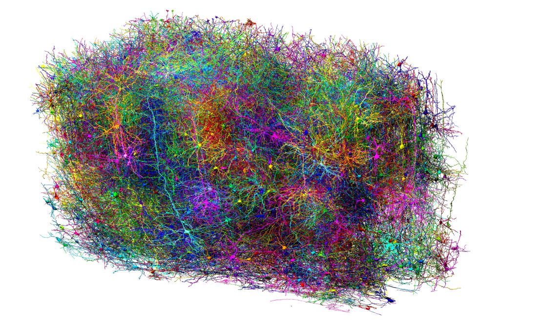
The card details the shape, function and activity of 84,000 neurons, branched structures that trigger messages in a long arm, called axone, then through more than 500 million synapses, as well as 200,000 brain cells. The tiny piece of fabric contained 3.4 miles (5.4 kilometers) of neural wiring – almost once and a half the length of the New York central park.
Work is the culmination of almost a decade of research by 150 scientists in 22 institutions led by the Allen Institute for Brain Science, the Baylor College of Medicine and the University of Princeton.
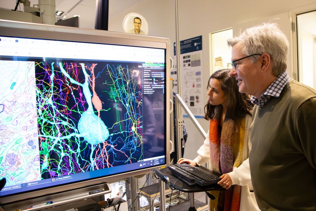
“A by-product of all this project shows us how beautiful the brain is,” said Dr. Forrest Collman, Associate Director of Data and Technology at the Allen Institute, in A video shared by the organization.
“The simple fact of looking at these neurons shows you their detail and their scale in a way that makes you appreciate the brain with a feeling of fear in the way when you look up, you know, say, on an image of a galaxy far, far,” he added.
The astonishing card represents only 1/500 of the full volume of the brain of a mouse, but the team ended up with 1.6 data petacts – an amazing quantity equivalent to 22 years of non -stop HD video, that the project, known as the Machine Intelligence of the Cortical Networks Program (Microns), has already published publicly.
Researchers described work in several articles Posted in La Revue Nature April 9.
To make the card, scientists from the Baylor College of Medicine of Houston began by using specialized microscopes to record brain activity in part of the cubic 1 fabric in the visual cortex of a laboratory mouse – where the animal treats what it sees – for a few days.
The researchers made sure that the mouse was awake and stimulated visually during the imagery by making the animal run on a treadmill and watching scenes of 10 seconds of various films, including “The Matrix” and “Mad Max: Fury Road”. The YouTube clips of extreme sports such as motocross, sledding and basic jump were also part of the rotation of visualization, according to a Press release from Princeton University.
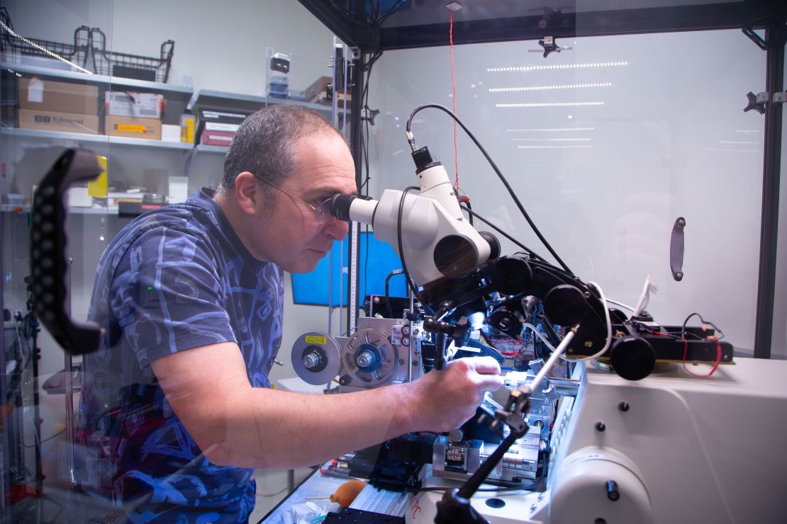
Then, after the euthanasia of the mouse, the researchers of the Allen of Seattle Institute took this same cube millimeter of brain and decided it in more than 28,000 layers, each 1/400 the width of a human hair, and took images of each slice along the way. They then rebuilt composite images.
“It took us about 12 days and 12 nights with the team that made changes 24 hours a day; and not because we cut it by hand, it is a machine that is automated,” said Dr. Nuno Maçarico Da Costa, an investigator associated with the Allen Institute.
“We had to be there to stop at any time if we thought we will lose more than a consecutive section.” If that happened, Da Costa said that experience should start from scratch, adding that the whole process was very “stressful”.
A team from Princeton University in New Jersey then deployed automatic learning and artificial intelligence tools to trace the outline of each neuron through the slices, coloring neurons to light them individually in a process called segmentation. The information generated by AI is validated or connected by scientists involved, a process that is still in progress.
The work has resulted in a unified view of what scientists call the brain of the “connectoma” mouse which shows how specific parts of the mouse brain are organized and offer an overview of the way different types of cells work together.
“Connectome is the beginning of the digital transformation of brain science,” said Dr. Sebastian Seung, Professor Evnin in neuroscience at Princeton University and IT teacher.
“With a few touchs, you can search for information and get the results in a few seconds. Some of this information would have taken an entire doctoral thesis to get before. And this is the power of digital transformation,” he said in a press release.
Brain cartography in this way had long been considered an impossible challenge. The molecular biologist Francis Crick, who won the Nobel Prize for describing the structure of DNA, could never be able to obtain such a detailed understanding of the brain.
“There is no point in asking for the impossible, as, say, the exact wiring scheme for a cubic millimeter of brain tissue and the way all its neurons shoot,” he wrote in Scientific American in 1979.
The “Connectome” mouse brain is based on similar work on even smaller creatures: the connection of the NEMATODE VER C. Elegans was completed in 2019 and scientists revealed a card of all The neurons of the brain of the fruit fly in 2024.
A cubic mouse brain millimeter is about 20 times larger than the full brain of the fruit fly, and much more complex, the researchers said. Nevertheless, the objective is to be able to map the entire mouse brain in the near future.
“I think that for the moment, the answer is no, it is not possible, but I think that everyone has really clear ideas on the way they could unravel these barriers. We hope in three or four years, we can say, yes, it is possible,” Collman told CNN.
However, he said that the cartography of the Connectome of the human brain in a similar synaptic resolution would be a radically more difficult business. “The human brain is another factor of about 1,500 larger than a mouse brain, and this brings a whole host … of technical and ethical barriers to that,” he said.
However, it could be possible to draw axons throughout the human brain, if not synaptic connections, added Dr. Clay Reid, a principal researcher in brain science at the Allen Institute.
“The prospect of reconstructing the whole human brain in terms of all connections is something for a distant future.”
The neocortex is particularly interesting to study, because this region of the brain is what distinguishes the brain from mammals from those of other vertebrates, said Dr. Mariela Petkova, research partner, and Dr Gregor Schuhknecht, postdoctoral scholarship holder, both in the Department of Molecular and Cellular Biology at the University of Harvard. Petkova and Schuhknecht were not involved in creating the mouse brain card.
“The researchers focused on this region, because it is generally considered to be the seat of superior cognition and plays a key role in sensory perception, language treatment, planning and decision -making”, ” They wrote in an article Posted alongside research.
“Remarkably, these apparently different functions are made possible by a plan which can be found, with some modifications, in all cortical zones and in all mammals.”
Laboratory mice are already widely used to understand human diseases, and a better understanding of the form and function of the brain of the mouse will present new possibilities to study human brain disorders such as Alzheimer’s disease, parkinson, autism and schizophrenia which involve disturbances in neural communication.
“If you have a broken radio and you have the circuit diagram, you will be better placed to repair it,” Da Costa said in a press release. “We describe a kind of Google card or plan of this sand grain. In the future, we can use it to compare cerebral wiring in a healthy mouse with brain wiring in a model of illness.”