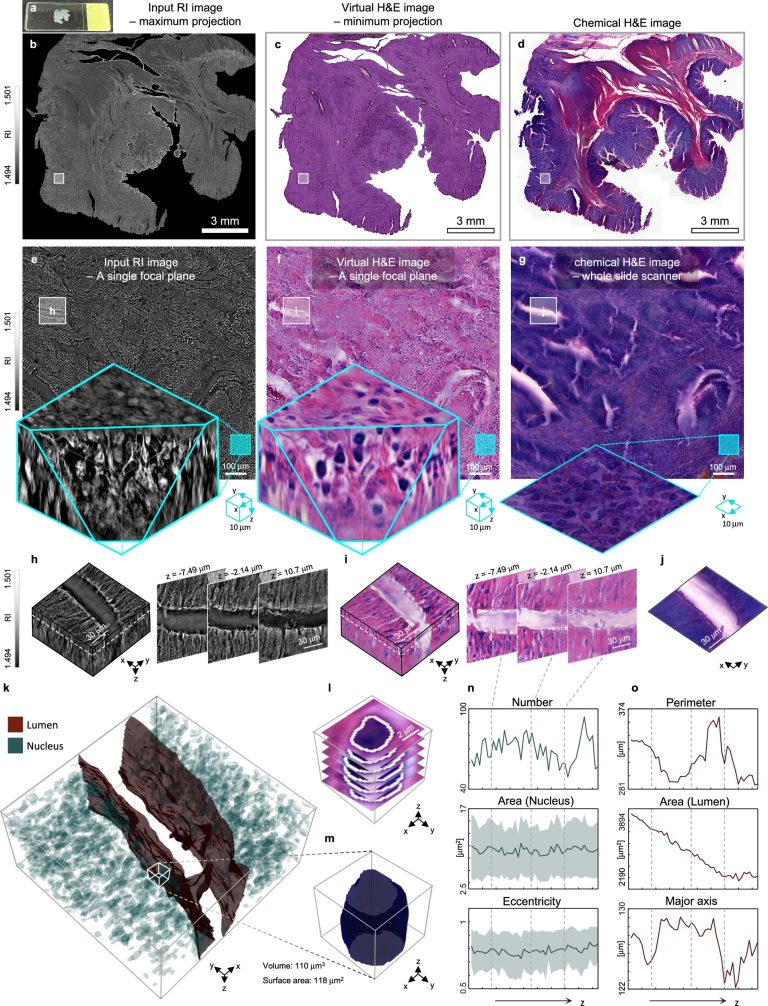
3D microanatomical rendering and quantitative analysis of a tissue tissue of colon cancer of 50 μm of 50 μm. Credit: Nature communications (2025). DOI: 10.1038 / S41467-025-59820-0
By moving beyond the traditional methods of observation of cancer fabrics in barely slices and stained, a collaborative international research team led by Kaist has successfully developed a new technology. This innovation uses advanced optical techniques combined with an in -depth learning algorithm based on artificial intelligence to create realistic and practically colorful 3D images of cancer tissue without needing an excisional biopsy. This breakthrough should pave the way for a new generation non -invasive pathological diagnosis.
A research team led by Professor Yongkeun Park of the Department of Physics, in collaboration with the team of Professor Su-Jin Shin at the Severance Hospital of the University of Yonsei, the team of Professor Tae Hyun Hwang at Mayo Clinic and the research team on Tomocube AI, developed an innovative technology capable of living in a living way the 3D structure of cancer fabrics Without separate coloring.
Research is published in the newspaper Nature communications.
For more than 200 years, conventional pathology has been based on the observation of cancer tissue under a microscope, a method that only shows specific transversal cuts of 3D cancer fabric. This has limited the ability to understand three -dimensional connections and spatial arrangements between cells.
To overcome this, the research team used holotomography (HT), an advanced optical technology, to measure the 3D refraction index information. They then joined a in -depth learning algorithm To successfully generate images of virtual hematoxyline and eosin – the most used coloring method to observe pathological tissues. Hematoxyline stains cell nuclei in blue and eosin stains pink cytoplasm.

Comparison of the conventional 3D tissue pathology procedure and 3D Virtual H & E virtual coloring technology proposed in this study. The traditional method requires preparing and sticking dozens of fabric blades, while the proposed technology can reduce the number of slides to 10 times and quickly generate H & E images without the coloring process. Credit: Korea Advanced Institute of Science and Technology (Kaist)
The research team has quantitatively demonstrated that the images generated by this technology are very similar to colorful fabric images. In addition, technology has presented coherent performance in various organs and tissues, proving its versatility and reliability as a new generation pathological analysis tool.
In addition, by validating the feasibility of this technology through joint research with hospitals and research institutions In Korea and the United States, using the holotomographic equipment of Tomocube, the team has demonstrated its large-scale adoption potential in the pathological research environments of the real world.
Professor Park said: “This research is a very significant realization which extends the unit of pathological analysis from 2D to 3D. It should be widely used in various biomedical research and clinical diagnoses, such as the analysis of the limits of cancer tumors and the spatial distribution of cells in the surrounding areas in the microtomor environment.”
More information:
Juyeon Park et al, revealing 3D microanatomical structures of thick cancer fabrics not marked using holotomography and h & e virtual coloring, Nature communications (2025). DOI: 10.1038 / S41467-025-59820-0
Quote: 3D virtual coloring technology allows non-invasive observation of cancer fabrics (2025, May 26) recovered on May 27, 2025 from https://medicalxpress.com/News/2025-05-3d-virtual-technology-inables-invasive.html
This document is subject to copyright. In addition to any fair program for private or research purposes, no part can be reproduced without written authorization. The content is provided only for information purposes.


